Upper Thigh Muscle Anatomy Mri : Mri Of The Thigh Detailed Anatomy Superior Part W Radiology : Anterior superior iliac spine insertion:
byAdmin•
0
Upper Thigh Muscle Anatomy Mri : Mri Of The Thigh Detailed Anatomy Superior Part W Radiology : Anterior superior iliac spine insertion:. Evidence translating to better performance and injury prevention. Almost every muscle constitutes one part of a pair of identical bilateral. Choose from 500 different sets of flashcards about thigh muscle anatomy on quizlet. There are several ways to do this. Muscle mri allows the identification of edema and fatty replacement of muscle tissue.
Anterior superior iliac spine insertion: Similar to fkrp distinguishing feature obturator externus & internus less involved than fkrp upper body common: We think this is the most useful anatomy picture that you need. Learn about thigh muscle anatomy with free interactive flashcards. There are around 650 skeletal muscles within the typical human body.
Hip And Thigh Muscles Anatomy And Functions Kenhub from thumbor.kenhub.com Whether it's to pass that big test, qualify for that big promotion or even master that cooking technique; The adductor muscles form the fleshy mass on the medial side of the thigh. Both the thigh and leg are divided into three separate compartments. Anterior superior iliac spine insertion: Human anatomy » musculoskeletal system » the muscles of the arm and hand. Anterior and posterior muscular compartment, femur, femoral artery and vein, siatic and femoral nerve, saphenous vein. Magnetic resonance imaging (mri) can be beneficial in identifying adductor brevis or adductor longus muscle atrophy which would indicate possible obturator nerve entrapment. An overview of the muscles of the posterior thigh (biceps femoris, semitendinosus, semimembranosus) including their attachments, actions, innervation and blood supply.
The gold standard for diagnosis of this condition is electromyography.
Human anatomy » musculoskeletal system » the muscles of the arm and hand. Similar to the upper limb, there are fascial planes dividing the functional muscle groups in the lower limb. The thigh has some of the body's largest muscles. The uppermost of the medial thigh muscles is the pectineus muscle. The gold standard for diagnosis of this condition is electromyography. Learn about thigh muscle anatomy with free interactive flashcards. Both the thigh and leg are divided into three separate compartments. Using mri as the reference method, muscle volume was predictedfrom anthropometry using a circular. As the name implies they adduct the thigh at the hip. Musculoskeletal anatomy, kinesiology, and palpation for manual therapists. The anterior femoral muscles (fig. Upper medial surface of the shaft of the tibia in front of the insertions of the gracilis and the semitendinosus nerve supply: The thigh is the area between the hip and the knee joint.
Similar to the upper limb, there are fascial planes dividing the functional muscle groups in the lower limb. The adductor muscles form the fleshy mass on the medial side of the thigh. It arises by tendinous fibers from the anterior superior iliac spine and the upper half of the notch below it. The muscular system is responsible for the movement of the human body. The thigh has some of the body's largest muscles.
Thigh Anatomy Mri Anatomy Drawing Diagram from www.researchgate.net There are several ways to do this. Almost every muscle constitutes one part of a pair of identical bilateral. Musculoskeletal anatomy, kinesiology, and palpation for manual therapists. Typical findings are edema, hematoma, and partial or complete muscles tears. Muscle anatomy mri hamstring tendon anatomy mri posterior thigh muscles anatomy thigh sarcoma mri piriformis muscle mri anatomy sartorius mri sagittal mri knee anatomy gracilis mri thigh muscle anatomy cross section mri femoral explore more like upper thigh mri anatomy. Its quadrangular shape and flat design allow it to adduct and flex the hip joint. Latissimus dorsi, serratus anterior, subscapularis uncommon: Choose from 500 different sets of flashcards about thigh muscle anatomy on quizlet.
It is part of the lower limb.
The muscles and fasciæ of the thigh. Using mri as the reference method, muscle volume was predictedfrom anthropometry using a circular. Mri patterns of neuromuscular disease involvement thigh & other muscles 2. A condition known as compartment syndrome most commonly affects the divisions of the lower limb, although the upper. Muscle mri can provide information that is complementary to clinical, histologic, genetic, and laboratory findings for the diagnosis of neuromuscular disease. The deltoid muscle is a rounded, triangular muscle located on the uppermost part of the arm and the top of the shoulder. Dummies helps everyone be more knowledgeable and confident in applying what they know. While the thigh muscles will be slip into the anterior, medial and posterior groups. We think this is the most useful anatomy picture that you need. There are around 650 skeletal muscles within the typical human body. Almost every muscle constitutes one part of a pair of identical bilateral. You can click the image to magnify if you cannot see clearly. Typical findings are edema, hematoma, and partial or complete muscles tears.
Discover the muscle anatomy of every muscle group in the human body. As the name implies they adduct the thigh at the hip. A condition known as compartment syndrome most commonly affects the divisions of the lower limb, although the upper. Whether it's to pass that big test, qualify for that big promotion or even master that cooking technique; The anterior femoral muscles (fig.
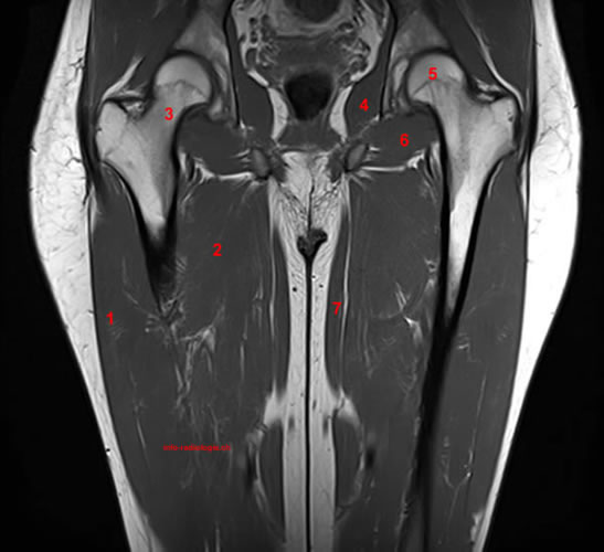
Mri Of The Thigh Detailed Anatomy Superior Part W Radiology from w-radiology.com Muscle mri allows the identification of edema and fatty replacement of muscle tissue. Muscles in the posterior compartment of the thigh. Imaging studies such as ultrasound, magnetic resonance imaging (mri), computed tomography (ct), herniography and laparoscopy can ↑ clinical gait, anatomy, and biomechanics of abdominal wall muscle. The muscles and fasciæ of the thigh. Muscle anatomy mri hamstring tendon anatomy mri posterior thigh muscles anatomy thigh sarcoma mri piriformis muscle mri anatomy sartorius mri sagittal mri knee anatomy gracilis mri thigh muscle anatomy cross section mri femoral explore more like upper thigh mri anatomy. Similar to fkrp distinguishing feature obturator externus & internus less involved than fkrp upper body common: Using mri as the reference method, muscle volume was predictedfrom anthropometry using a circular. The muscles of the torso, examined in the previous chapter, include a few that attach directly into the upper arm and help move the humerus at the shoulder joint.
Anatomy of the thigh :
Magnetic resonance imaging (mri) can be beneficial in identifying adductor brevis or adductor longus muscle atrophy which would indicate possible obturator nerve entrapment. The uppermost of the medial thigh muscles is the pectineus muscle. Upper medial surface of the shaft of the tibia in front of the insertions of the gracilis and the semitendinosus nerve supply: Find the best weight lifting exercises that target each muscle or groups of you can click the links in the image, or the links below the image to find out more information on any muscle group. Choose from 500 different sets of flashcards about thigh muscle anatomy on quizlet. You can click the image to magnify if you cannot see clearly. This is a table of skeletal muscles of the human anatomy. Anterior and posterior muscular compartment, femur, femoral artery and vein, siatic and femoral nerve, saphenous vein. The muscular system is responsible for the movement of the human body. There are several ways to do this. Almost all muscles cross at least one joint (moveable connection between two bones) and cause an action across that joint. Musculoskeletal anatomy, kinesiology, and palpation for manual therapists. Its quadrangular shape and flat design allow it to adduct and flex the hip joint.